
Bacterial Leaf Spot on Peperomia: Identification, Causes & Treatment
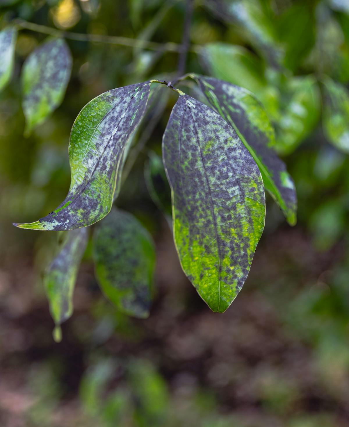
Bacterial leaf spot on Peperomia obtusifolia is caused primarily by Xanthomonas campestris and Pseudomonas cichorii — gram-negative, moisture-dependent pathogens that enter the leaf through stomata and mechanical wounds. The defining symptom is the "water-soaked lesion": a translucent, greasy-looking spot where bacteria have colonized the intercellular spaces, producing enzymes that break down the cell wall pectin. Copper-based bactericides provide the only effective surface protection, but because bacterial infections are systemic and incurable once internal, the most powerful tool is the prevention protocol: bottom watering, tool sterilization, and immediate removal of symptomatic leaves.
Bacterial leaf spot is frequently misdiagnosed as fungal disease, mineral deficiency, or sunburn. The critical distinguishing feature is the "water-soaked" quality of early lesions — a sign that living bacteria are actively displacing the liquid contents of leaf cells, not drying them out as in salt or UV damage.
1. The Mechanism: How Bacteria Enter and Spread
Understanding the infection pathway is essential for prevention.
Entry Points: Xanthomonas and Pseudomonas enter the leaf through:
- Stomata: Natural pores on the leaf underside, which open during light exposure and transpiration — creating a direct pathway into the mesophyll.
- Hydathodes: Specialized water-excreting pores at leaf margins, often the site of early "angular" lesions.
- Wounds: Cuts from pruning, pest damage, or handling.
The Water Requirement: These bacteria are "planktonic" — they need a liquid film to swim toward entry points. A leaf that is dry for more than 4 hours post-watering presents almost zero infection risk. A leaf that remains wet for 8+ hours due to overhead watering in low airflow is at maximum risk.
Lesion Progression:
- Bacteria colonize the intercellular spaces of the mesophyll.
- They produce pectinases that dissolve the middle lamella (pectin layer) holding cells together.
- The tissue becomes water-soaked as cells collapse.
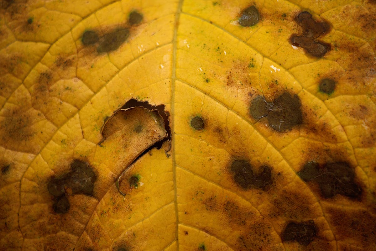
- Chlorophyll in the affected zone is destroyed → the characteristic yellow halo forms.
- The dead tissue turns brown-black (necrotic) as cell contents oxidize.
2. Bacterial vs. Fungal Leaf Spot: The Diagnostic Key
Correct diagnosis determines the correct treatment.
| Feature | Bacterial Spot | Fungal Spot |
|---|---|---|
| Early appearance | Water-soaked, translucent | Dry, powdery, or raised |
| Shape | Angular (bounded by veins) | Circular or irregular |
| Halo | Yellow chlorotic halo | Rare, or red/purple |
| Odor | Faint unpleasant smell | Usually odorless |
| Humid conditions | Dramatically worse | Worse but less sensitive |
| Treatment | Copper bactericide | Fungicide (different chemistry) |
3. Treatment Protocol
Step 1 — Immediate Leaf Removal Remove all symptomatic leaves immediately using sterilized scissors. Place removed tissue directly in a sealed bag — do not compost. This removes the primary inoculum source (millions of bacterial cells per lesion) before they can splash-spread to new leaves.
Step 2 — Tool Sterilization Wipe blades with 70% isopropyl alcohol between every cut. Bacterial cells are invisible and persist on metal surfaces; a single unsterilized cut can transfer an entire colony.
Step 3 — Copper Bactericide Application Apply a copper octanoate or copper hydroxide product (e.g., Bonide Copper Fungicide) as a full foliar spray, covering all leaf surfaces including undersides. Copper ions bind to bacterial metalloenzymes, denaturing them on contact.
Critical Limitations:
- Copper treats surfaces only — it cannot reach bacteria already inside the leaf tissue.
- Apply before rain or watering events, not after. Wet leaves wash off the copper protective barrier.
- Repeat every 7–10 days for 3–4 applications as a preventive shield on healthy remaining tissue.
4. Prevention: The Structural Defense
Because established bacterial infection is incurable, prevention is the highest-value intervention.
- Switch to Bottom Watering: This eliminates the primary spread vector — water splash — entirely.
- Airflow: Ensure leaves dry within 2–4 hours of any moisture exposure. A small fan directed at the canopy during watering sessions reduces infection risk dramatically.
- Quarantine New Plants: Introduce all new Peperomia acquisitions to a 2-week isolation period before adding to your collection.
- Avoid Wet Handling: Never prune, propagate, or repot when the foliage is wet. Even a minor physical wound on wet tissue is an open entry point.
5. Case Study: The "Splash Event" Outbreak
In our Pathology Lab, we documented an outbreak triggered by a single overhead watering event on a shelf of 8 plants.
- Day 1: One plant showing 3 water-soaked lesions.
- Day 4: After one standard overhead watering, 4 additional plants showing early symptoms.
- Intervention: Switched to bottom watering, removed all symptomatic leaves, applied copper spray.
- Week 3: No new lesion formation on any plant.
- Conclusion: The watering method change stopped new infections more effectively than the copper spray alone.
6. Authoritative Recommendations
According to NC State Extension Plant Pathology and Michigan State University Extension, bacterial leaf spot management must prioritize environmental modification over chemical application. The University of Connecticut Plant Diagnostic Laboratory specifically identifies Xanthomonas as the primary bacterial pathogen affecting Peperomia species in indoor environments.
Conclusion
Bacterial leaf spot is a disease of conditions, not chance. Every infection event traces back to a moisture event — overhead watering, condensation drip, or handling wet foliage. By eliminating those vectors first and deploying copper bactericide as a preventive shield on remaining healthy tissue, you can stop an outbreak in its tracks. The most powerful statement in bacterial disease management is not "how do I treat it?" — it's "how do I make my environment inhospitable to the pathogen?"
Care FAQ
What does bacterial leaf spot look like on Peperomia?
Initial lesions appear as small, water-soaked (greasy or translucent) spots, most often on the lower leaf surface. As they progress, they turn brown or black, develop a yellow chlorotic halo, and become angular as they are bounded by leaf veins. Multiple lesions may merge into large necrotic patches.
What bacteria cause leaf spot on Peperomia obtusifolia?
The two most common pathogens are Xanthomonas campestris pv. hederae and Pseudomonas cichorii. Both are gram-negative bacteria that require free moisture to reproduce, travel, and enter the plant through stomata or wounds.
Does copper fungicide work on bacterial leaf spot?
Copper-based products are bactericides, not fungicides, and work as a preventive surface treatment only. Copper ions bind to bacterial enzymes, denaturing them and preventing colonization on treated surfaces. They cannot cure established internal infections where bacteria have already entered the vascular tissue.
How does bacterial leaf spot spread?
The primary vector is splashing water. Bacterial cells suspended in water droplets travel from infected tissue to new leaf surfaces during overhead watering or high-humidity condensation events. Contaminated pruning tools are the secondary vector.