White Spots on Peperomia Obtusifolia: Edema, Pests, or Normal Anatomy?
White spots on Peperomia obtusifolia trigger an immediate assumption: pests. Sometimes that is correct. More often it is not. Five distinct things produce white markings on this species — one pest, two physiological disorders, one fungus, and one entirely normal anatomical feature — and the correct response ranges from "isolate and treat immediately" to "do nothing, this is healthy biology." Reaching for pesticide before identifying which of the five you are looking at wastes product, stresses the plant, and leaves the actual cause uncorrected.
The direct answer: White spots on P. obtusifolia are caused by mealybugs (cottony, wipe off), oedema (hard corky bumps on the underside), powdery mildew (dry dusty coating), hard-water mineral deposits (chalky surface crust), or cystoliths (calcium carbonate crystals that are a normal part of the leaf). A single 10-second wipe test, combined with the spot's texture and location, separates all five and dictates the correct intervention.
Quick Diagnostic Table
| Appearance | Location | Wipes Off? | Cause | Action |
|---|---|---|---|---|
| Cottony, fuzzy, sticky | Leaf axils, midrib, undersides | Yes — smears | Mealybugs | Isolate + treat |
| Fine stippling, faint webbing | Underside, spreading | Partial | Spider mites | Isolate + treat |
| Dry, powdery, dusty film | Upper surface, spreading | Yes — dusts off | Powdery mildew | Isolate + treat |
| Hard, raised, corky bumps | Underside, no halo | No — part of leaf | Oedema | Adjust watering/airflow |
| Chalky crust | Tips, near stomata | Scrapes with nail | Mineral deposits | Switch water source |
| Flush or recessed dots, permanent | Either surface | No — under skin | Cystoliths (normal) | None — leave |
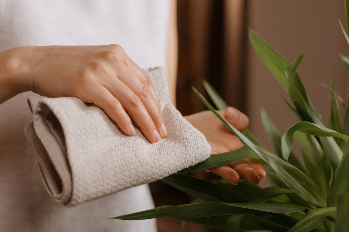
1. The 10-Second Wipe Test
Before any treatment, run the single diagnostic that separates the five causes into three families. Take a damp microfibre cloth or a cotton swab dipped in 70% isopropyl alcohol and gently wipe one white spot.
- It smears, lifts off, or leaves residue on the cloth. The marking sits on the leaf surface — a mealybug colony (sticky, cottony), spider mite debris (fine, with webbing), or powdery mildew (dry, dusty). These are the biological and contagious causes.
- It does not move, but sits as a hard raised bump or a chalky crust. The marking is bonded to the surface — oedema scar tissue (corky, underside) or hard-water mineral deposit (chalky, scrapes with a fingernail).
- It does not move and is flush with or recessed into the leaf. The marking is inside the leaf — a cystolith, part of the plant's own architecture.
The wipe test is the fork in the diagnostic tree. Everything below follows from which of the three results you got.
2. Mealybugs — The Pest That Wipes Off
Mealybugs (Pseudococcus longispinus and relatives) present as white, cottony masses concentrated in the leaf axils, along the midrib, and on the undersides — the sheltered points where they pierce the phloem to feed. Clusters of fine white egg sacs accompany an established infestation. They feel soft and sticky, and they smear when wiped — the defining contrast with oedema and cystoliths.
The mechanism is sap extraction. Mealybugs drain phloem, weakening the plant and excreting honeydew that becomes a substrate for sooty mould. Spider mites (Tetranychus urticae) produce a related but distinct white signature — fine stippling and faint webbing rather than cottony masses, with outbreaks driven by RH below 40%.
The treatment: Isolate the plant immediately. Wipe colonies off with a cotton swab dipped in 70% isopropyl alcohol, which dissolves their protective wax coat on contact. Follow with a neem oil solution (2 ml per litre of water plus a drop of mild soap as emulsifier) applied to all surfaces and undersides every 5–7 days for three cycles to interrupt the reproductive cycle. The full protocol is in the mealybug identification and treatment guide; for the webbing-and-stippling variant, see the spider mite guide.
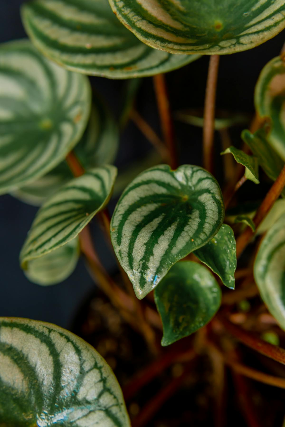
3. Oedema — Hard Corky Bumps That Will Not Move
Oedema is not an infection — it is a pressure-rupture event, and it is the most common non-pest cause of white-to-tan markings on this species. P. obtusifolia stores water in its semi-succulent leaves. When the substrate is wet and the surrounding air is cool, humid, or stagnant, the roots absorb water faster than the stomata can release it through transpiration. Internal hydrostatic pressure builds until epidermal cells on the leaf underside physically rupture; the ruptured cells oxidise and suberise into hard, corky, raised scabs that read as white, tan, or brown bumps.
A recurring scenario: a specimen on a windowsill is watered, then experiences a cool, still night — radiator off, low airflow. Within 24–72 hours, water-soaked spots appear on the underside of the lower leaves and harden into corky scars. The damage is permanent in the affected tissue; new growth under stable conditions emerges clean.
The treatment: The existing scars do not reverse. Prevent new ones by raising the transpiration rate — increase airflow with a gentle fan, allow the top 2–3 cm of substrate to dry fully before watering (every 10–14 days in summer, 21–28 days in winter for a 12 cm pot), and use a free-draining mix with at least 30% inorganic content. The cellular-level mechanism and full prevention protocol are in the oedema guide.
4. Powdery Mildew — The Dry White Film
Powdery mildew is a fungal infection presenting as a dry, dusty, white-to-grey coating that starts as small patches on the upper leaf surface and spreads to a film. Unlike mealybug fluff, it is not sticky and not concentrated in axils; unlike oedema, it sits on top of the leaf and dusts off onto the cloth in the wipe test.
The fungus colonises leaf surfaces in warm, humid, stagnant air — the same low-airflow conditions that drive oedema, which is why the two sometimes appear together. It does not require standing water to germinate, distinguishing it from bacterial and many fungal leaf-spot pathogens.
The treatment: Isolate the plant. Improve airflow to disrupt the still boundary layer the fungus needs. Wipe off the visible coating, then treat with a neem oil or potassium-bicarbonate solution on a 7-day cycle until new growth emerges clean. The full protocol is in the powdery mildew treatment guide.
5. Mineral Deposits — The Chalky Hard-Water Crust
Hard tap water carries dissolved calcium, magnesium, and bicarbonates. When water evaporates from the leaf surface — especially after misting, or where droplets sit on the foliage — these minerals are left behind as a chalky white crust, typically near the leaf tips and over the stomata. It scrapes off with a fingernail, leaving a powdery residue, which separates it from both the smear of a pest and the immovable bump of oedema.
This is a cosmetic and water-source issue, not a health threat to the plant. The same mineral load, when taken up through the roots and accumulated in the substrate, drives the separate problem of leaf-tip salt burn — see the white crust and salt buildup guide for the substrate side.
The treatment: Wipe deposits off with a cloth dampened with distilled or filtered water. Switch routine watering to low-mineral water — rainwater, distilled, or filtered — to prevent recurrence. The mechanism and water-source comparison are in the tap water sensitivity guide.
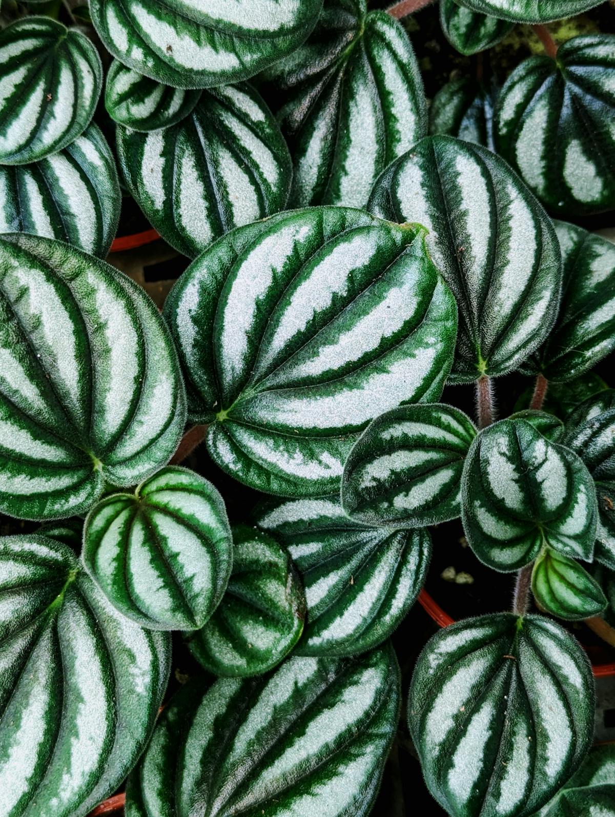
6. Cystoliths — When the White Spots Are Healthy Anatomy
The most-misdiagnosed white markings on P. obtusifolia are not a problem at all. The standard reaction — "white dots, it must be a pest, spray it" — is wrong here, and the pesticide does nothing because there is no organism. These are cystoliths: crystals of calcium carbonate that the plant forms inside specialised epidermal cells called lithocysts. They are a normal, defining feature of the Piperaceae family's leaf anatomy, present in healthy specimens.
The mechanism is mineral management. P. obtusifolia often absorbs more calcium than it needs; rather than let it reach toxic concentration in the sap, the plant sequesters it as solid calcium carbonate inside lithocysts. The crystals also scatter light into the deeper mesophyll, helping the chloroplasts below the surface capture photons. They are flush with or slightly recessed into the leaf, they do not move under the wipe test, and they are permanent for the life of the leaf because they are structural, not superficial.
The treatment: None. Cystoliths require no intervention and are a sign of normal metabolism. If their density bothers you aesthetically, watering with low-mineral water reduces cystolith formation in new growth — but existing crystals remain, and there is no reason on plant-health grounds to act at all.
When to Do Nothing
Three of the five causes need no intervention beyond an environmental adjustment, and one needs nothing whatsoever:
- Cystoliths: leave entirely. This is healthy anatomy.
- Mineral deposits: cosmetic only; wipe and switch water source at your convenience.
- Oedema: the existing scars are permanent; only the watering and airflow need correcting to prevent new ones.
Only mealybugs, spider mites, and powdery mildew demand prompt, active treatment, because only those three are living and contagious. The discipline of this species is restraint: identify with the wipe test first, and intervene only where a living organism is confirmed.
Recommended Products
These are the tools the treatments above call for. Specificity matters more than brand.
- 70% isopropyl alcohol — dissolves the mealybug wax coat on contact; the first-line spot treatment applied by cotton swab.
- Cotton swabs — for precise alcohol application to mealybug colonies in leaf axils without wetting the whole leaf.
- Neem oil concentrate — the follow-up treatment for mealybugs, mites, and powdery mildew over the pest life-cycle.
- Digital hygrometer — confirms whether RH and airflow sit in the band that prevents oedema and mildew (
40–60% RH, moving air). - Distilled water or filter pitcher — low-mineral water to stop hard-water deposits and reduce new cystolith formation.
Sources and Related Reading
- Wikipedia — Cystolith (the calcium-carbonate-in-lithocyst anatomy)
- Wikipedia — Oedema in plants (the pressure-rupture disorder)
- NC State Extension — Peperomia obtusifolia (species profile; mealybug and pest references)
Internal mechanism references on this site:
- Mealybug identification and treatment — the pest cause in depth.
- Spider mite identification and treatment — the stippling-and-webbing variant.
- Oedema / blistered leaves guide — the corky-bump cause in depth.
- Powdery mildew treatment — the dry-film fungal cause.
- White crust and salt buildup — the substrate side of mineral accumulation.
- Tap water sensitivity — water-source comparison for mineral deposits and cystoliths.
- Leaf anatomy and succulence — where cystoliths and the mesophyll sit in the leaf.
Summary
White spots on P. obtusifolia are a five-way differential, not a single problem. The wipe test sorts them in ten seconds: anything that smears or dusts off is a living, contagious cause (mealybugs, spider mites, powdery mildew) and needs prompt isolation and treatment; anything that stays put is either a permanent oedema scar, a wipeable mineral crust, or a cystolith — a healthy calcium structure that needs nothing. Identify before intervening, and reserve the pesticide for the one case in five that actually warrants it.
Care FAQ
What are the tiny white dots on my Peperomia leaves?
Run the wipe test first. If they smear or lift off, they are mealybugs (cottony, sticky) or powdery mildew (dry, dusty). If they are hard corky bumps on the underside that will not move, they are oedema scars. If they are a chalky crust near the tips, they are hard-water mineral deposits. If they are flush with or recessed into the leaf surface and permanent, they are cystoliths — natural calcium carbonate crystals that are harmless and require no treatment.
Are white spots on Peperomia a sign of pests?
Not always. Of the five common causes, only one — mealybugs (or spider mites) — is a pest. The other four are physiological oedema, powdery mildew (a fungus), hard-water mineral deposits, and cystoliths (normal anatomy). Treating a non-pest cause with pesticide wastes product and stresses the plant. Identify before you intervene.
Do white cystolith spots need treatment?
No. Cystoliths are calcium carbonate crystals the plant forms inside specialised epidermal cells called lithocysts — a normal feature of the Piperaceae family. They are flush with the leaf surface, permanent, and harmless; they cannot be wiped off because they are part of the leaf architecture. If their number bothers you aesthetically, watering with low-mineral water reduces new cystolith formation in future growth, but existing ones remain.
How do I tell oedema from mealybugs?
Texture and removability. Mealybugs are soft, cottony, and sticky, sit on the leaf surface (often in leaf axils and along the midrib), and wipe off with a cotton swab dipped in 70% isopropyl alcohol. Oedema scars are hard, corky, raised, sit almost always on the leaf underside, and cannot be removed without tearing the leaf — they are suberised scar tissue, not an organism.
Will white spots spread to my other plants?
Only the biological causes spread. Mealybugs, spider mites, and powdery mildew are contagious and warrant immediate isolation. Oedema, mineral deposits, and cystoliths are non-contagious — they are reactions or structures specific to the individual plant and its environment, and pose no risk to neighbouring specimens.